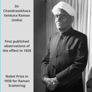
Raman spectroscopy has transformed analytical chemistry, providing insights into molecular composition and structure. This article explains 20 types of Raman spectroscopy, from Surface-Enhanced Raman Spectroscopy (SERS) to Time-Resolved Raman Spectroscopy to Micro Raman Spectroscopy.
The focus here is to understand the distinction from the traditional Raman setup and highlight their applications.
The Basics of Raman Spectroscopy
Raman spectroscopy is an analytical technique that involves the interaction of light with a sample and analyzing the scattered light to gain information about its molecular composition.
When light interacts with molecules, most of it scatters unchanged (Rayleigh scattering), but a small fraction undergoes a phenomenon called Raman scattering. This scattered light contains valuable information about the vibrations and energy levels of the molecules.
Raman spectroscopy measures the Raman shift, which is the difference in energy between the scattered light and the incident light. It produces two types of lines: Stokes lines (lower energy) and Anti-Stokes lines (higher energy). By analyzing these lines, scientists can identify and study the molecules present in the sample.
Types of Raman Spectroscopy
Regardless of the specific type used, Raman spectroscopy relies on the detection and analysis of the Raman scattered light, which is sensitive to molecular properties and provides insights into chemical composition, structure, and dynamics.
While the specialized types of Raman spectroscopy may incorporate additional elements such as enhanced signals, specific excitation wavelengths, or advanced imaging techniques, they are all built upon the fundamental principles of Raman scattering and the analysis of molecular vibrations.
1. Confocal Raman Spectroscopy
Confocal Raman spectroscopy combines Raman spectroscopy with confocal microscopy, allowing for spatially resolved Raman spectra. It provides detailed information about the chemical composition and structure of specific regions within a sample.
Confocal Raman spectroscopy offers improved spatial resolution and the ability to perform 3D imaging, distinguishing it from traditional Raman spectroscopy.
It is useful in materials characterization, biological imaging, and studying heterogeneous samples.
2. Surface-Enhanced Raman Spectroscopy (SERS)
SERS utilizes nanostructured metallic surfaces to enhance the Raman signal, enabling sensitive detection of trace analytes.
It offers significantly higher sensitivity compared to traditional Raman spectroscopy, making it valuable for trace analysis, single-molecule detection, and studying interfaces and surfaces.
Surface-Enhanced Raman Spectroscopy finds applications in biosensing, environmental monitoring, and forensic analysis.
3. Coherent Anti-Stokes Raman Spectroscopy (CARS)
It is a nonlinear type of Raman spectroscopy technique that uses two laser beams to probe molecular vibrations.
CARS provides higher sensitivity and faster acquisition times compared to traditional Raman spectroscopy.
It is particularly useful for studying complex mixtures, biological samples, and real-time imaging of dynamic processes, finding applications in medical diagnostics and chemical analysis.
4. Resonance Raman Spectroscopy
This type involves exciting the sample with a laser wavelength matching the electronic transition energy of the molecule.
Resonance Raman spectroscopy enhances the Raman signal and provides information about electronic states.
It is valuable for studying electronic and vibrational coupling, excited states, and chromophores in various fields such as materials science, catalysis, and biological research.
5. Surface-Enhanced Spatially Offset Raman Spectroscopy (SESORS)
SESORS combines SERS and spatially offset Raman spectroscopy to collect Raman spectra from beneath optically scattering or absorbing materials.
It enables analysis through scattering or opaque layers, making it useful for non-destructive characterization of materials, forensics, and biomedical applications.
6. Tip-Enhanced Raman Spectroscopy (TERS)
This type of Raman Spectroscopy involves scanning a sharp metallic or dielectric tip across a sample surface to enhance the Raman signal at the nanoscale.
TERS enables high-resolution chemical imaging and analysis with spatial resolution beyond the diffraction limit of light.
It finds applications in nanomaterials characterization, surface plasmonics, and studying molecular interactions at the nanoscale.
7. Time-Resolved Raman Spectroscopy
It studies molecular vibration dynamics using ultrafast laser pulses to excite and probe the sample on a picosecond or femtosecond timescale.
Time-resolved Raman spectroscopy provides insights into transient species, reaction kinetics, and energy transfer processes.
It is valuable for investigating photochemical reactions, excited states, and dynamic processes in materials, chemistry, and biological systems.
8. Polarized Raman Spectroscopy
This type utilizes polarized incident light and measures the polarization dependence of the scattered light to study molecular orientation and anisotropy.
Polarized Raman spectroscopy provides information about molecular symmetry, crystallographic properties, and orientation of molecules in various materials, such as liquid crystals, polymers, and crystals.
9. Micro-Raman Spectroscopy
It performs Raman spectroscopy with a microscope to obtain spatially resolved information at a microscopic scale.
Micro-Raman spectroscopy enables the characterization of small particles, thin films, biological samples, and microelectronic devices.
It is useful in materials science, forensics, and biological research.
10. Stimulated Raman Scattering (SRS)
This type of Raman spectroscopy involves the interaction of a pump beam and a Stokes beam to amplify the Raman signal.
SRS provides higher sensitivity and faster acquisition compared to traditional Raman spectroscopy, allowing for label-free imaging and quantitative analysis.
It finds applications in biomedical imaging, chemical analysis, and studying live cells and tissues.
11. Spatially Offset Raman Spectroscopy (SORS)
SORS collects Raman spectra from subsurface layers by utilizing the spatial separation of the incident and collected light.
It enables non-destructive analysis of opaque or turbid samples, such as pharmaceutical tablets, packaging materials, and biological tissues.
It is valuable in quality control, pharmaceutical analysis, and security screening.
12. Nonlinear Raman Spectroscopy
This type includes CARS, stimulated Raman gain (SRG), and stimulated Raman loss (SRL) techniques, which exploit nonlinear interactions between light and molecules to enhance Raman signals.
Nonlinear Raman spectroscopy provides higher sensitivity, improved spatial resolution, and faster acquisition times compared to traditional Raman spectroscopy.
It is useful for studying complex materials, biological systems, and chemical dynamics.
13. Transmission Raman Spectroscopy (TRS)
TRS collects Raman spectra from the transmitted light through a sample, enabling analysis of bulk materials and pharmaceutical tablets without the need for extensive sample preparation.
It offers improved depth profiling capabilities and is useful for quantitative analysis, pharmaceutical quality control, and non-destructive testing.
14. Coherent Raman Scattering (CRS)
Coherent Raman scattering is a type of Raman spectroscopy that encompasses CARS, coherent Stokes Raman scattering (CSRS), and coherent anti-Stokes rotational Raman spectroscopy (CARSRR).
CRS techniques utilize coherent interactions between laser beams and molecules to amplify Raman signals. CRS provides higher sensitivity, faster acquisition, and 3D imaging capabilities compared to traditional Raman spectroscopy.
It finds applications in biomedical imaging, materials analysis, and studying complex mixtures.
15. Dispersive Raman Spectroscopy
This type involves dispersing the Raman scattered light using a grating or prism and detecting the intensity at different wavelengths to obtain a Raman spectrum.
Dispersive Raman spectroscopy provides detailed spectral information and is commonly used for routine analysis, identification of unknown compounds, and structural analysis.
16. Ultraviolet Raman Spectroscopy
It utilizes ultraviolet (UV) light as the excitation source, which enhances the Raman scattering from certain molecules.
UV Raman spectroscopy offers increased sensitivity to specific compounds, electronic transitions, and surface analysis, making it useful for studying materials with strong fluorescence or inorganic materials.
17. Near-Infrared Raman Spectroscopy
This type involves using near-infrared light for excitation, which reduces fluorescence interference and provides better penetration depth.
Near-infrared Raman spectroscopy enables analysis of highly scattering samples, such as tissues and powders, and is utilized in pharmaceutical analysis, biomedical diagnostics, and food quality control.
18. Inelastic Neutron Scattering (INS)
It uses neutrons instead of photons to probe the vibrational modes of molecules.
INS provides complementary information to Raman spectroscopy, particularly for hydrogen-containing compounds and lighter elements.
It is valuable for studying materials in extreme environments, catalysts, and hydrogen storage materials.
19. Raman Optical Activity (ROA)
This type measures the difference in Raman scattering intensity for left- and right-circularly polarized incident light to determine the chiral properties of molecules.
ROA provides information about molecular structure, conformation, and chirality, making it valuable in studying biomolecules, pharmaceuticals, and materials with chiral properties.
20. Resonance SERS (SERRS)
Resonance SERS combines the enhancements of SERS with resonance Raman spectroscopy, utilizing a resonant excitation wavelength.
It provides enhanced sensitivity and selectivity for analytes with specific electronic transitions, enabling trace-level detection and identification in complex samples.
It finds applications in biosensing, environmental analysis, and forensic science.
